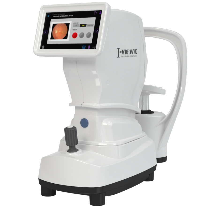
I-VIEW100
AI-assisted fundus camera for first-line screening of three blinding diseases.
- Type
- Non-mydriatic retinal camera
- Resolution
- 13 MP
- Field of view
- 50°
- Min. pupil
- 3.5 mm
- Focus range
- −20 D to +20 D
- Display
- 7" touch screen
What it is
I-VIEW100 is a fundus camera with an onboard AI algorithm for primary screening of retinal disease. It is built to be operated by non-ophthalmologist medical staff: it finds the eye, focuses and shoots automatically, then returns a traffic-light risk indication within one second of capture.
System components — 380 (W) × 450 (D) × 400 (H) mm, 16 kg. Motorised chinrest with 35 mm travel, base movement 42 mm forward/back, 88 mm left/right, 42 mm up/down. USB 3.0. AC 100–240 V 50/60 Hz.
Indications
- First-line screening for glaucoma
- First-line screening for diabetic retinopathy
- First-line screening for macular degeneration
Specifications & configurations
Manufacturer documentation for I-VIEW100. Click any image to open it full size.
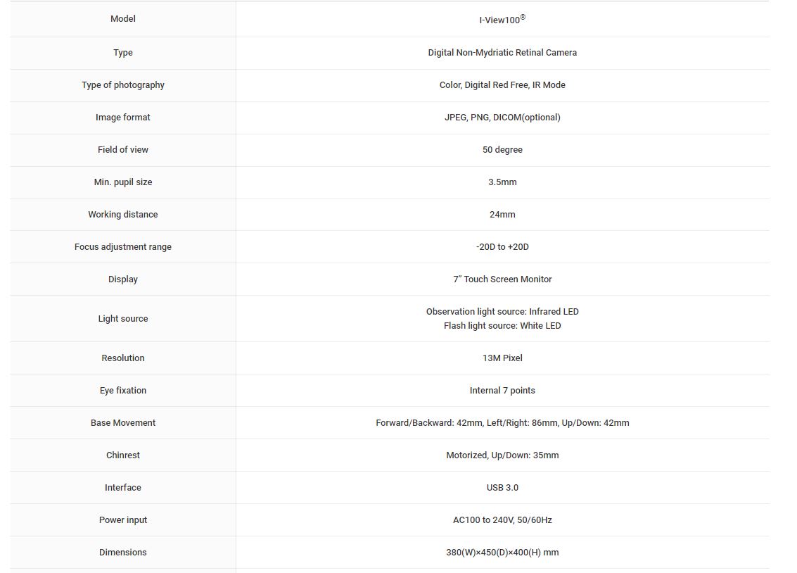
Key features
AI algorithm on board
Performs first screening for three blinding diseases and presents the outcome as a red, yellow or green indicator within one second of image capture — a format that non-specialist staff can act on immediately.
Auto shooting mode
Auto tracking, auto focusing and auto shot remove the operator skill barrier that normally limits where fundus photography can be deployed.
Designed for non-specialists
A 7 inch touch-screen panel with joystick control, intended for use by medical staff who are not ophthalmologists.
Compact footprint
380 × 450 × 400 mm and 16 kg — small and light enough for a primary-care room, a mobile screening unit or a community programme.
Four imaging modes and DICOM output
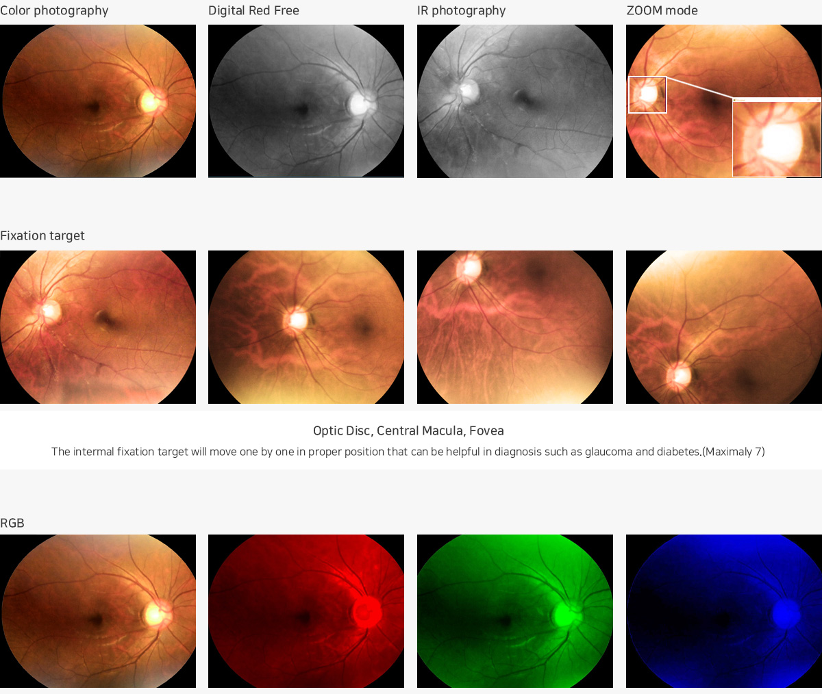
Colour, digital red-free and infrared photography plus zoom mode, at 13 MP with a 50° field of view. Images export as JPEG or PNG, with DICOM available as an option for integration into existing imaging systems.
Seven-point internal fixation
The internal fixation target steps through seven positions so the optic disc, central macula and fovea can each be brought into position — the coverage needed for glaucoma and diabetic retinopathy screening.
The underlying principle
Screening value depends on throughput, not on single-image quality. Automating alignment, focus and capture — then triaging the result to three states rather than producing a diagnosis — is what makes retinal screening viable outside specialist ophthalmology.
Clearance & regulatory position
Bio-Med USA distributes FDA-cleared medical, aesthetic, surgical and dental devices. Clearance numbers, indications-for-use statements and the current regulatory position for I-VIEW100 in your market are provided on request.